|
Diseases of the liver is a large parenchymal organ and gland situated in the RUQ structurally characterised by being the largest gland in the body and one of two organs with a portal circulation and functioning as the metabolic warehouse
Inflammation

Radiation Hepatitis – Hyperemia
|
| The straight line and the hyperemia are characteristis features radiation induced change
22975 Courtesy Ashley Davidoff MD |
| Liver capsule |
| (Image courtesy of Ashley Davidoff M.D.) |
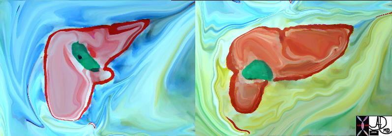
Normal Liver and Cirrhosis |
|
Parts of the liver changing in size reflecting disease. The first image reflects a normal liver In the second image the left lobe is relatively large while the right lobe is small. These finding are characteristic of alcoholic cirrhosis. Note the shape of the surface of the liver has also changed from being smooth to being nodular and the presence of aciteds reflects a combination of increased portal pressure and low proteins.
18135.800 46136 Davidoff MD |
|
Normal Liver above Cirrhotic Liver below |
| This diagram reflects the large left lobe of the liver in cirrhosis and the small right lobe. The caudate lob is not depicted. 42649c01 Davidoff art |
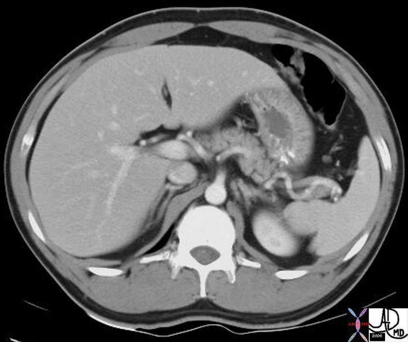
Abscess Complicating Traumatic Infarction of the Liver
|
| 24010 liver fx air fx air fluid level loculated air free air dx hepatic abscess following traumatic injury CTscan Courtesy Ashley Davidoff MD |

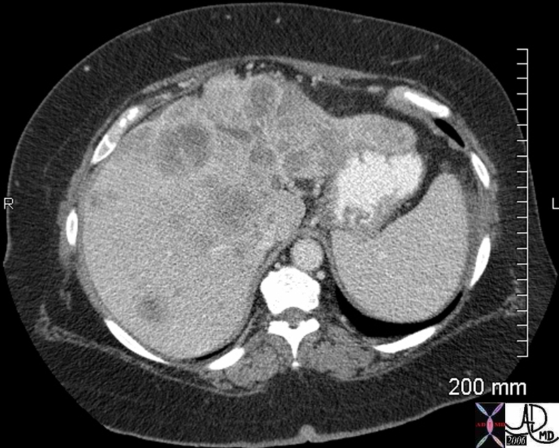
HCC small
|
| 48369c03 40 male with hepatitis B liver fx hypervascular lesion seen in early arterial phase only with rapid wasout dx HCC hepatoma hepatocellular carcinoma capillary hemangioma characterisation characterization blood flow CTscan MRI Courtesy Ashley DAvidoff MD |
 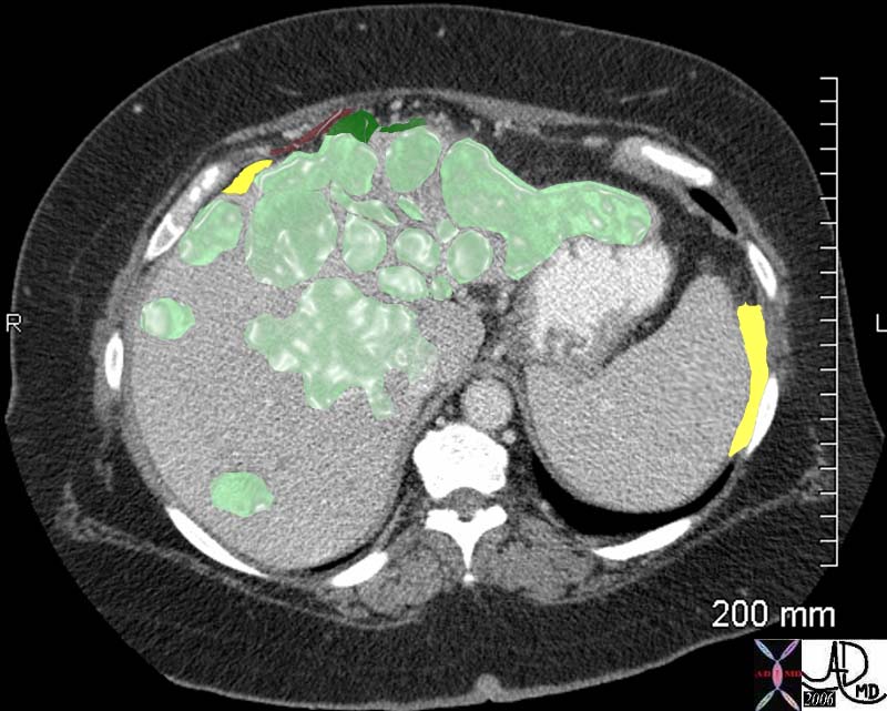
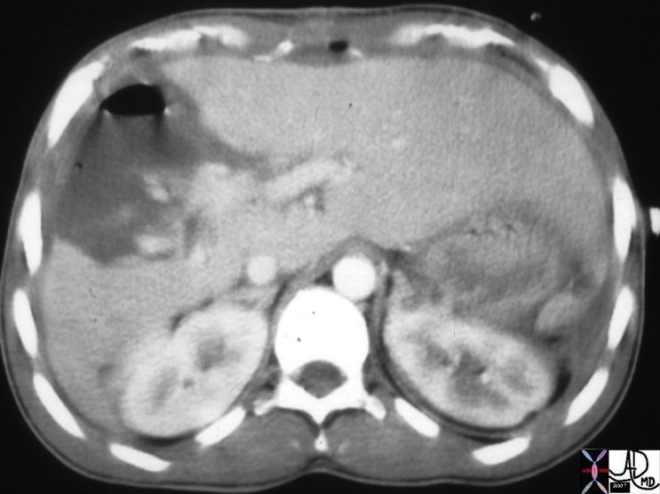
Colon Carcinoma with Metastases to the Liver
|
| The abdominal CT is from a middle aged female with right sided discomfort. The image reveals multiple space occupying lesions (light green) in the liver. The patient was found to have a primary colonic carcinoma. The capsule of the liver is involved by tumor extending to the surface of the liver and deforming the surface (dark green). The disease is also close to the diaphragm (maroon). The involvement of the liver capsule and possibly the right hemidiaphragm is likely the cause of the patients pain. There is also a small amount of ascites present (yellow) either as a response to involvement of the liver capsule or the early development of malignant ascites.
45051 45051b04
middle aged female with right sided discomfort liver diaphragm fx enlarged hepatic enlargement hepatomegaly fx hepatic masses shrunken left lobe fx abdominal ascites dx colonic carcinoma with hepatic metastasis (metastatic liver disease) metastases with diaphragmatic and renal displacement from the large liver ascites probably malignant note hepatic capsule is involved abdominal CTscan of the abdomen Courtesy Ashley Davidoff MD 45049 45050 45051 45052 45053 45054 |
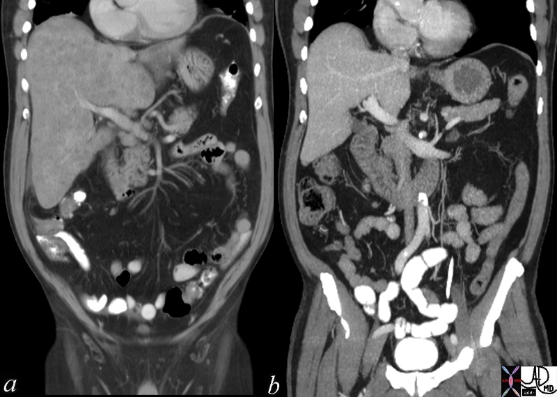
Chemotherapy – Before and 6 weeks After – Metastatic Small Cell Lung Carcinoma |
| 70248c01 liver metastattic small lung carcinoma with diffuse metatstattic disease to the liver )hepatic metastases metastasis before and after treatment 6weeks post chemotherapy successful result size change character change CTscan Davidoff MD 5star |
Metabolic Liver Disease
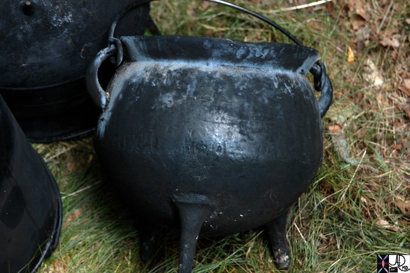
Iron Pots – Bantu Beer – Acquired Iron Overload Disease
|
| 84108p.800 tripod iron pots Bantu beer acquired hemosiderosis acquired iron overload Davidoff photography Davidoff MD |
|