Length: 15-17.5 cm
Transverse: 20-22.5 cm
Antero-posterior: 10-12.5 cm
Weight: 1.2-1.8 kg
Volume:
The adult liver has a craniocaudal span of 15-17.5 cm, a transverse diameter of 20-22.5 cm, and an antero-posterior depth of 10-12.5 cm. It weighs between 1.2-1.6 kg (3.5 lbs.)
The liver is the largest gland in the body, accounting for about 2% of the body weight in adults and 5% in infants. It is also the largest abdominal organ. It usually weighs more in males than females.
The right lobe is usually six times as large as the left.
Since the left lobe is variably sized and is commonly congenitally small or absent.
A transverse diameter 20-22.5 cm, is therefore very variable.
Size
In the male, the liver weighs between 1.4 to 1.6 kg, and in the female, between 1.2 to 1.4 kg. Its greatest horizontal measurement is from 20 to 22.5 cm. Vertically, near the right surface, it measures about 15 to 17.5 cm., while its greatest antero-posterior diameter is on a level with the upper end of the right kidney, and measures 10 to 12.5 cm.
The liver
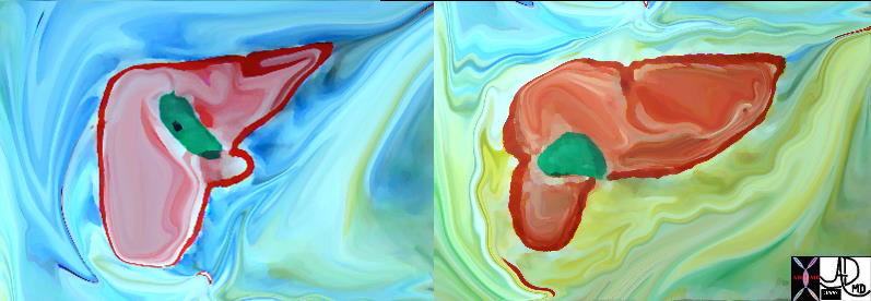
In this color overlay the liver is seen in relation to other upper abdominal organs. Note its dominance in the upper abdomen. (Image Courtesy of Ashley Davidoff M.D.)

The linear measurements of the liver are not easy to apply in the clinical context. Sometimes the right lobe will reach all the way down to the iliac crest as a normal variant called a Riedel’s lobe. The size and shape of the left lobe is a better indicator of the size of the liver. Using planimetry and density conforming techniques, the volume and hence the size of the liver is more accurately assessed. This method is not yet automated and therefore only performed under special circumstances such as in the screening of candidates for liver transplantation. The normal volume of the liver is between 1200 and 1500 cc’s.
|
Left lobe |
|
The left lobe has been manually outlined and shaded in this liver donor patient. This process has to be repeated at all levels for the volume of the left lobe to be determined. It is essential therefore to know and understand the boundaries of the liver lobes and segments to accurately outline and measure them. (Image Courtesy of Ashley Davidoff M.D.) |

A case of a larger than normal liver.
|
Left lobe |
|
This image shows a large left lobe of the liver – compare the its size to a normal sized left lobe below. (Image courtesy of Ashley Davidoff M.D.) |

A case of a smaller than normal liver.
|
Lobular shaped liver |
|
This liver has suffered the consequences of cirrhosis, in which liver tissue is replaced by scar tissue resulting in an overall shrinkage of the liver. |

Size Applied Anatomy
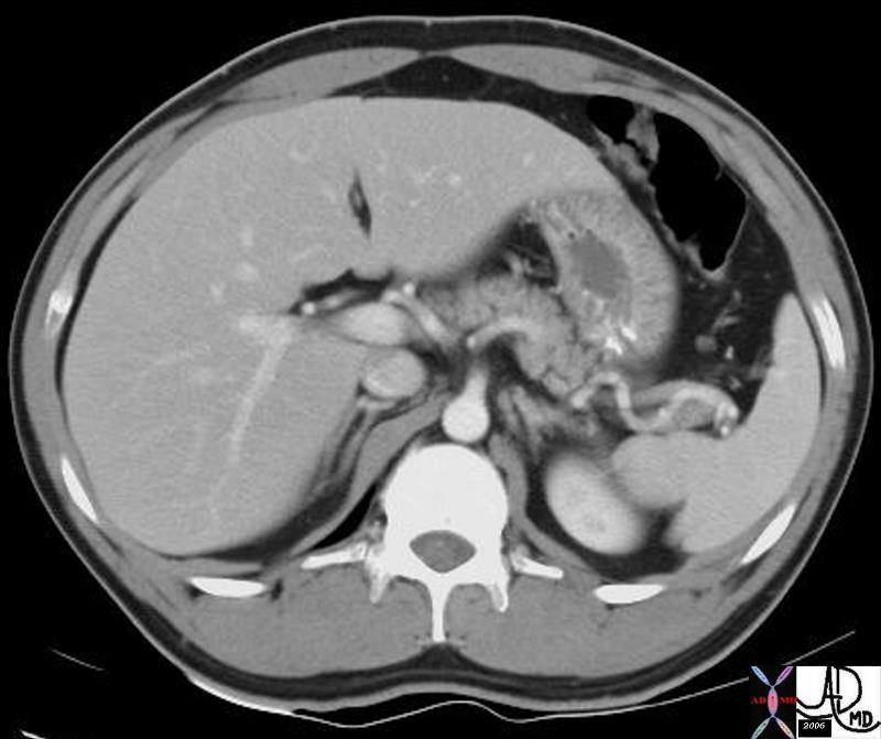
This image shows a small lateral segment (II and III) of the left lobe. This condition is called congenital hypoplasia of the left lobe, implying that the person was born with this anatomic variant. It has no functional significance.
|
Congenital hypoplasia of the left lobe. |
|
Congenital hypoplasia of the left lobe. (Image Courtesy of Ashley Davidoff M.D.) |

In this patient, part of the anterior segment (V)of the liver is missing and the colon pokes its head through the missing right lobe segment. This condition is also congenital and has no clinical significance.
|
Congenital hypoplasia of the right lobe. |
|
Congenital hypoplasia of segment 4 of the right lobe. (Image Courtesy of Ashley Davidoff M.D.) |

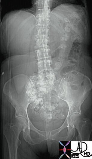
Riedels lobe
Riedel’s lobe is a variant an inferior extension of the right lobe of the liver occurring more commonly in women. It is a normal anatomic variant and its presence changes both the size and shape of the right lobe of the liver.
|
Riedel’s lobe |
|
This plain film of the abdomen from the scout of a CT scan shows the classical findings of a Ridel’s lobe with an elongated right lobe of the liver that has vertiical length that exceeds the horizontal breadth disprportionaetely (“the lean and hungry look”) 25255 (Image courtesy of Ashley Davidoff M.D.) |
|
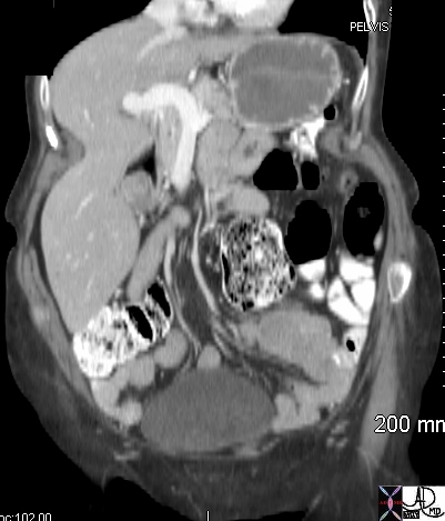
Riedel’sobe |
|
The coronal image from a CT scan shows an example of a Riedel’s lobe . Note also that this image reveals the malleable nature of the liver which is easily deformed by the indentation caused by the muscle and the ribs of the lateral abdominal wall. (Image courtesy of Ashley Davidoff M.D.) 44963 |
|
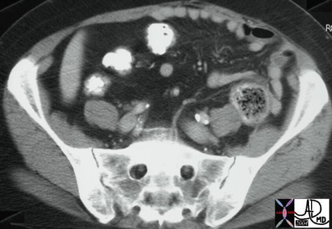
Tip of liver in RLQ – Riedels lobe |
|
The tip of the liver in this image is noted in the right lower quadrant.. The shape of this inferior extension is narrow and almost tongue like in its shape. This gracile appearance of the of the liver is characteristic of the Riedel’s lobe. (Image courtesy of Ashley Davidoff M.D.) 25258 |
Normal Liver and Cirrhosis |
|
Parts of the liver changing in size reflecting disease. The first image reflects a normal liver In the second image the left lobe is relatively large while the right lobe is small. These finding are characteristic of alcoholic cirrhosis. Note the shape of the surface of the liver has also changed from being smooth to being nodular and the presence of aciteds reflects a combination of increased portal pressure and low proteins. 18135.800 46136 Davidoff MD |
|
Normal Liver above Cirrhotic Liver below |
| This diagram reflects the large left lobe of the liver in cirrhosis and the small right lobe. The caudate lob is not depicted. 42649c01 Davidoff art |